Share
A fetal cervical teratoma is an extremely rare kind of tumor that develops most often in the thyroid gland, and one that can be life-threatening. The word “teratoma” comes from the Greek word for “monster,” and it’s perfectly appropriate. Formed from errant germ cells, a teratoma is made up of all kinds of tissues, including hair, teeth, muscle, and bone, all jumbled together in a grotesque mass. Although they are not cancerous, they can grow very quickly and to great size—as large as a softball or even a soccer ball—distending the fetus’s neck to an extraordinary degree. It’s that size and growth rate that pose the danger. The tumor can completely cut off the baby’s airway, and it can require so much blood to feed its growth that it can threaten the baby’s heart. As long as the baby is in the womb, the blockage of the airway isn’t an issue, because the baby isn’t breathing air yet. But when the baby is born, that blockage is life-threatening: the baby has only a few minutes to live without oxygen, and no oxygen can get to the lungs.
Up until the last 30 years or so, that condition was almost always fatal. But in the past few decades, doctors have developed new techniques and approaches that allow them to perform surgery on fetuses while they are still in the womb. And these teratomas are among the conditions they can now treat.
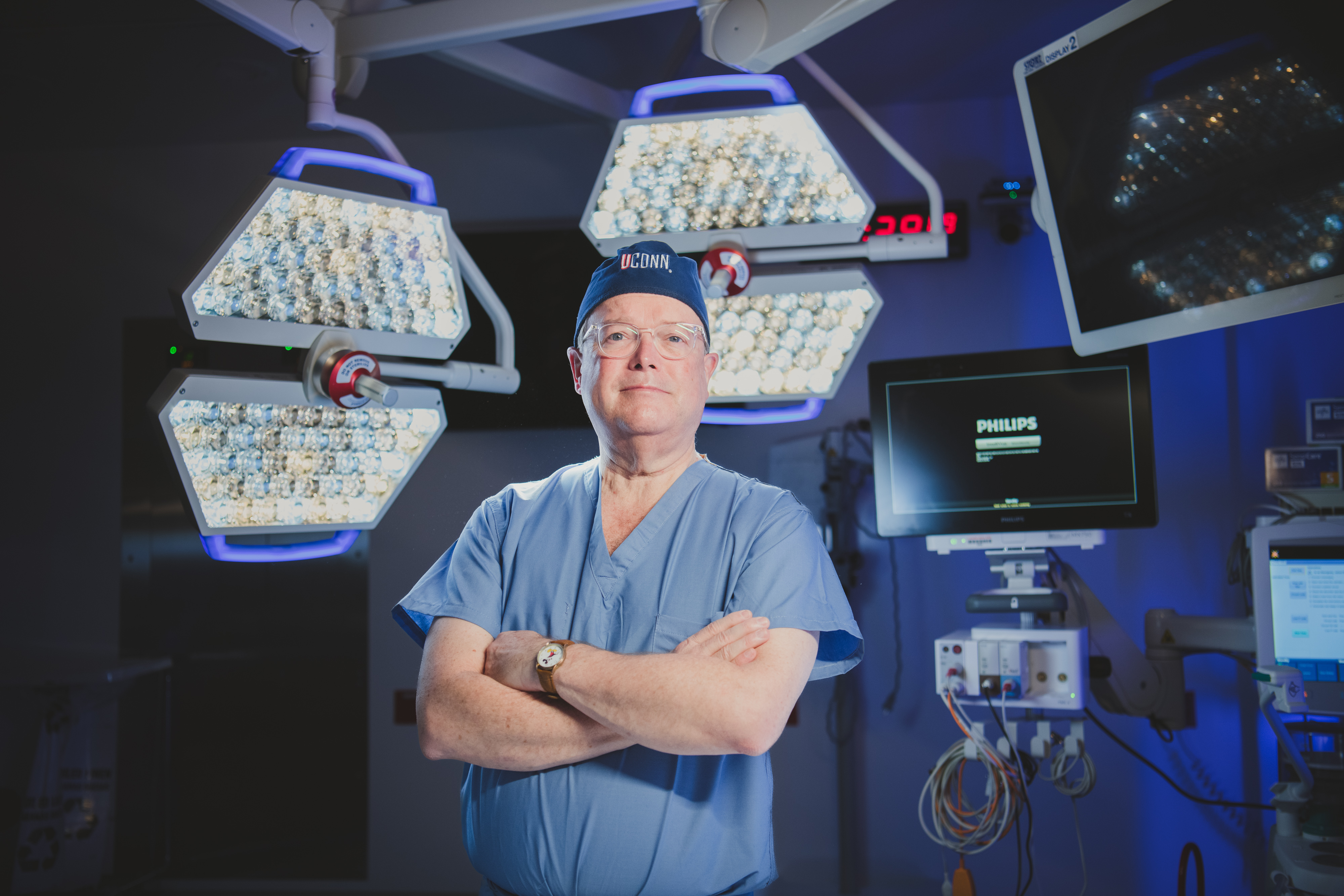
This cutting-edge surgery is just one of an amazing range of procedures that will be available at the Fetal Care Center, an exciting part of Connecticut Children’s expansion plan. The Center will be headed by Timothy M. Crombleholme, MD, one of the leading lights of this medical specialty. He has helped establish some of the top fetal care centers in the country and has co-authored more than 300 peer-reviewed articles, book chapters and the textbook Fetology: Diagnosis and Management of the Fetal Patient, which is widely considered the gold standard in the field.

A World of Solutions
In addition to cervical teratomas, the Fetal Care Center will be able to address a wide range of fetal problems with an equally wide range of tools and techniques. “There are, for example, ultrasound-guided procedures,” Dr. Crombleholme says. “Using a needle, we can do intrauterine transfusions. We can tap fluid spaces. We can do amnio infusions. We can put a laser fiber into a blood vessel feeding a lung tumor and photocoagulate the vessel to shrink that tumor. We can devascularize teratomas that have these enormous vessels that feed them. We can do ultrasound-guided procedures with a radio frequency device, which uses radio frequency to generate heat that can coagulate the feeding vessel.”
But that’s not all. There's a condition called twin reversed arterial perfusion sequence. In this situation, a normal multiple-fetus pregnancy has gone awry, and one of the twins is nothing more than a fetus-shaped mass with no head, no heart, and no nervous system. It is connected to the healthy fetus and draws blood from it to such a degree that the healthy baby can die from heart distress. Dr. Crombleholme addresses this condition by using ultrasound to guide a 19-guage needle to the cord that connects the healthy fetus and the malformed one. The needle has a special tip that uses radio frequencies to coagulate the cord. The unviable fetus dwindles, and the healthy fetus no longer loses nutrients and blood. “We have a 98 percent survival with that approach,” Dr. Crombleholme says.
A Womb with a View
“We can also do open fetal surgery,” Dr. Crombleholme says, “most commonly for repairing spina bifida, but also taking out masses like pericardial or mediastinal tumors. We can do amnioports for amnio infusion, and a whole host of EXIT procedures.”
EXIT stands for ex-utero intrapartum treatment, one of the most complex procedures the Fetal Care Center will perform and the one that addresses cervical teratomas—the tumor in a fetus’s neck that chokes off the airway. The surgeon makes an incision in the mother’s abdomen and uterus, then pulls the fetus partway out and performs the necessary surgery while the fetus is still receiving oxygen and nourishment from the placenta. When the procedure is done, the baby is fully delivered. In other open fetal surgeries, the fetus is returned to the womb, incisions sutured and the pregnancy continues.
The idea is audacious, and actually accomplishing it requires overcoming multiple hurdles, achieving precise timing and maintaining intensive monitoring and adjustment. Because the procedure involves two patients, the mother and the baby, it requires a wider-than-usual range of specialists, with as many as 40 people in the room. “You have myself,” Dr. Crombleholme says, “and a maternal-fetal-medicine specialist, another fetal surgeon, a cardiologist doing continuous echocardiographic monitoring plus a scrub team—at least one, sometimes two scrub teams—an obstetric anesthesiologist, a pediatric anesthesiologist, the neonatal resuscitation team, and, if you're going to continue operating after the baby's delivered, a whole other OR team and another setup.” That’s why a Fetal Care Center requires a larger-than-average operating room, to accommodate all those people and equipment necessary for the procedure.
The EXIT procedure requires balancing competing needs and liabilities. For example, the mother needs to be deeply anesthetized, initially with an IV anesthetic and then, once the baby is partially delivered, with inhaled anesthetic. That second anesthetic is crucial, because it causes the uterus to relax. That, in turn, means that placental gas exchange can continue for the fetus during the procedure. But there are two problems: first, cutting into a relaxed uterus causes massive hemorrhaging. So the surgeons use a specially designed uterine stapling device that stops the blood flow at the incision. The second problem is anesthetizing two patients. The mother must be deeply anesthetized to provide uterine relaxation and the fetus must be anesthetized to undergo surgery. There is 20- to 30-minute lag time before the fetus achieves the same depth of anesthesia as the mother. To bridge this lag time, the anesthesiologist gives the fetus an intramuscular injection of a cocktail of fentanyl, atropine and vecuronium.
Because there are so many moving parts and interacting systems involved, Dr. Crombleholme needs to be aware of all the monitoring systems. “The anesthesiology team needs to know what the relaxation of the uterus is like,” he says. “The echocardiographer needs to know that fetal heart rate is normal and the cardiac state is normal. And the whole team needs to know that on an ongoing basis, the maternal-fetal-medicine specialist is monitoring the uterine wall tone, while preventing delivery of the baby. And then the operative team are starting an IV, placing a pulse oximeter and giving the cocktail injection and then starting on the fetus’s airway. Now, with the airway, you have to come loaded for bear. You need direct laryngoscopy and flexible and direct bronchoscopy to see what’s there. You might need to open the fetal neck to release the strap muscles to take pressure off the trachea, which may allow the endotracheal tube to pass the obstruction. We might need to approach the airway through the chest. You need to have all those tools in your toolbox.”
The reason the monitoring is so important is because the margin for error is so slim. For example, if the mother’s uterus begins to tense and leave that relaxed state, it may not be reversible, and the surgeons may have to stop the EXIT procedure and immediately deliver the baby with the neck mass still in place. “Then,” Dr. Crombleholme says, “you're scrambling to open the airway with a baby who's just been born. As soon as they clamp the umbilical cord, that baby is becoming hypoxic and you've got less than five minutes before the baby has irreparable brain injury, or it goes into full arrest.”
EXIT procedures are used for several kinds of life-threatening conditions. For some of these conditions, like highly vascular prenatally diagnosed sacrococcygeal teratoma, the EXIT procedure has changed the prognosis from near 100 percent mortality to over 90 percent survival.
Just Because We Can Doesn’t Mean We Should
Just because a baby might benefit from fetal surgery, that doesn’t necessarily mean they will get fetal surgery. “We’ve had some parents who, for psychosocial reasons, we’ve had to say ‘no’ to,” Dr. Crombleholme says. “They were not good candidates because of a psychiatric illness that we thought would put them at great risk, that the stress of going through this—which is no walk in the park—could tip them over the edge. That's why we have psychologists evaluate these moms.” Even when the mother has no psychosocial issues, there can still be a need for counseling. The mother is often very enthusiastic to have the procedure done, while her partner may be more concerned about the risks to the mother. In those cases, the team needs to help the mother to understand all of the possible risks to her and complications for the child. For example, one of the conditions that Dr. Crombleholme has recently added to the list of treatments is fetal renal (kidney) failure. In and of itself, kidney failure for a fetus is a moot point, since the fetus doesn’t need to use their kidneys’ filtering function. Everything is filtered through the placenta, and the mother excretes the waste via her kidneys. For a fetus, the real problem in renal failure is the lungs. The lungs cannot develop if there’s no amniotic fluid, and after about 16 weeks’ gestation, all of the amniotic fluid comes from urine output by the baby. If the baby’s kidneys are not working properly, the urine cannot get to the bladder and out into the amniotic sac. And without that fluid, the lungs cannot develop, and these babies die a pulmonary death at delivery, not living long enough for the renal failure to be an issue.
“In the past,” Dr. Crombleholme says, “if a baby were to present with bladder outlet obstruction due to posterior urethral valves, and the kidneys are dysplastic and the bladder doesn't really refill because the kidneys are no longer making much urine, then that mother would've been told, ’There’s nothing you can do; go home. The baby will either die in utero, or, if you do make it to delivery, the baby will pass within an hour.’”
That was before. Now, Dr. Crombleholme uses a needle or an amniotic port to inject sterile IV fluid into the amniotic sac, so the lungs have fluid supporting them, and they develop naturally. Those babies no longer need to die. That doesn’t, however, mean that they are in the clear. Once the baby is born, the kidney problem that was moot in utero becomes a major issue. That baby will need dialysis within days and then a kidney transplant. And that, in turn, means a complicated conversation with the parents.
“A large part of our job is educating parents about what these options are and the risks and the benefits,” Dr. Crombleholme says. “It's not clear for many families that this makes sense. This may not be a good option. For something like fetal renal failure, we insist that they meet with neonatology to talk about having a baby in the NICU for protracted periods of time; a pediatric nephrologist, so they can hear about how complicated it is caring for a baby with renal failure from birth; a transplant surgeon about the limitations of getting from birth on dialysis to getting to a kidney transplant and that you're basically exchanging one chronic disease for another; sometimes the pediatric urologist, because either there is no bladder, as in bilateral renal agenesis, or the bladder is really destroyed in the case of bladder outlet obstruction; and then a psychologist to make sure that we're not going to push this family over the edge by the stress of undergoing this process. And we refuse to agree to treat them until they've been through this process. After hearing from all of these various subspecialists, the parents think that, ‘Yes, we think we can do this,’ then we go ahead.”
Care that Starts Before the Beginning and Continues After the End
That pre-procedure education is the beginning of what Dr. Crombleholme calls vertically integrated care, which includes caring for the mother and child through diagnosis and treatment through delivery, through the Neonatal Intensive Care Unit, and then during long-term follow up, all with the same team of doctors and clinicians.
“If we can reduce the stress that these parents experience when their baby's in the nursery,” Dr. Crombleholme says, “then they can be a parent as opposed to a passive bystander with the nurses and the doctors hovered around the baby. They can be the mom and the dad. My experience has been that it makes an enormous difference in the neurodevelopmental outcome of these children, if their parents can be parents from the time the baby is born, rather than being so intimidated by the medical setting, the baby's going to have a better physical outcome.”
All of the conditions treated in the Fetal Care Center are rare to one degree or another. But there are some conditions that are far from rare and, as yet, have no treatment available. One of those is fetal growth restriction. As the name suggests, the fetus does not grow at the typical rate and is at risk of a whole host of medical problems. Many babies with this condition are stillborn. In fact, this condition is the second leading cause of mortality just before or after birth. But Dr. Crombleholme, in addition to his work in the operating room, is also a researcher, and he is working on a method of gene therapy that holds great promise to reverse this condition.
“It affects 7 percent of all pregnancies and there is no treatment for it,” Dr. Crombleholme says. “The fetus is not growing and they're doing very poorly. They get delivered severely premature with all the attendant risks of severe prematurity. In my basic research in mouse, rat and rabbit models, we’ve been able to do placental gene therapy to correct the growth restriction. We can expand the vascularity of the placenta and completely reverse the growth restriction in utero. And growth restriction in these animals has the same characteristics as human growth restriction. Not only is that animal a normal size when it’s born, but the non-treated growth-restricted fetuses, when they become adult animals, are obese, diabetic, and have a hypertensive cardiomyopathy. None of those things happen with the animals treated in utero. This is something that we’re very excited about, and we think that is just over the horizon, that we’re going to be able to make the transition from treating animal models to treating growth-restricted babies.”
Latest Articles

Three Students Awarded Isidore Wise Scholarships