Whoops!

This website no longer supports this web browser. Use one of these browsers to get the best possible experience.

In the 1950s and ’60s, one of the popular themes for science fiction movies was mad scientists growing human brains in the lab. At the time, the notion of growing any part of a human in the lab was both unimaginably advanced and entirely disturbing (the story never turned out well for the scientist or anyone else). Today, we are actually living that reality, with scientists who are anything but mad, and their results are poised to transform medicine and the lives of countless children.

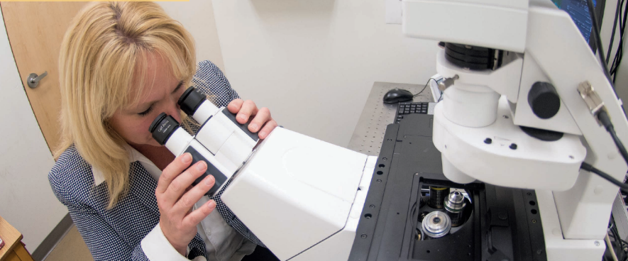
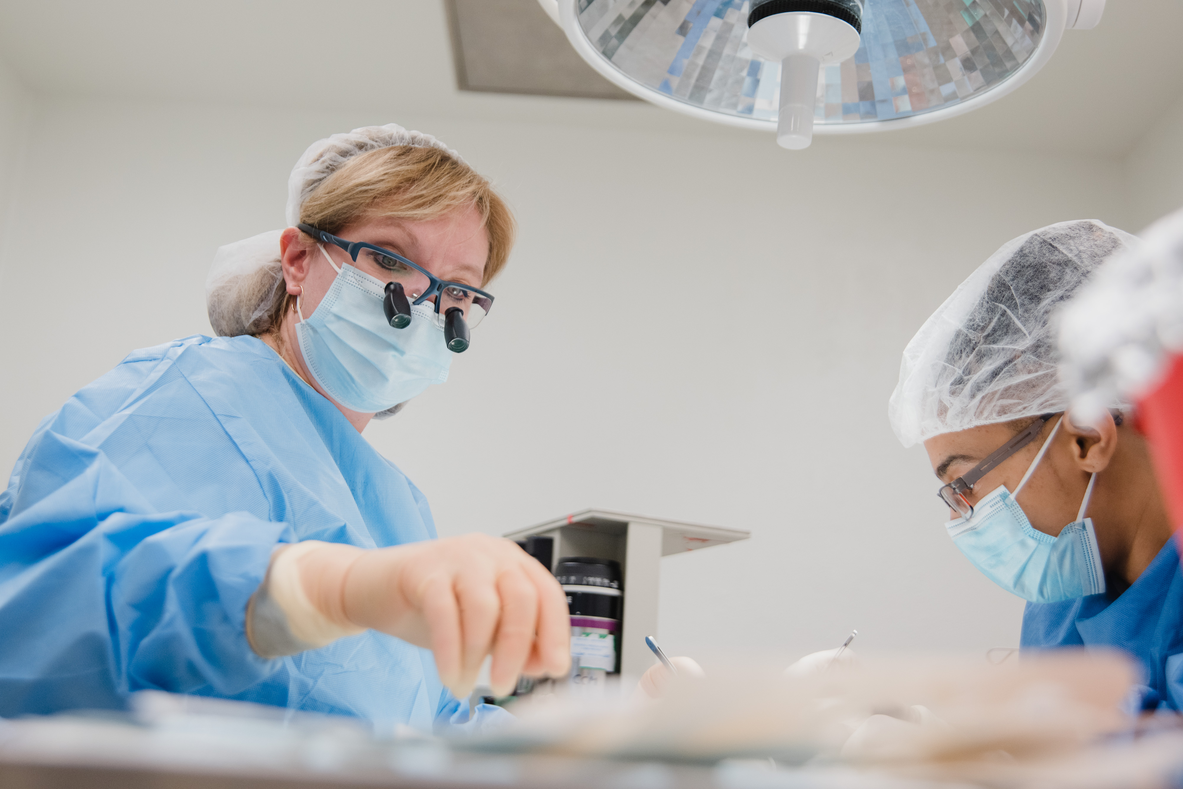
Christine Finck, MD, is Connecticut Children’s Surgeon in Chief and also the head of the Division of Pediatric General and Thoracic Surgery. She currently holds the Peter J. Deckers Endowed Chair in Pediatric Surgery. In addition to all her work as a surgeon and an administrator, Dr. Finck is conducting some of the most advanced research anywhere: she is working in regenerative medicine, using stem cells and a 3D bioprinter to grow human organs.

Bioprinting is a Reality

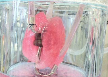
A 3D bioprinter is similar to a regular 3D printer in that it builds a three-dimensional object layer by layer. But where a conventional 3D printer might use plastic or ceramic material to build up those layers, a bioprinter can use cells as ink. And the object it creates is organic tissue.

The first work Dr. Finck undertook in this area was the esophagus, the tube that connects the mouth and the stomach. Human babies and animals are sometimes born with an incomplete esophagus, just two opposing stubs that don’t meet. In this situation, food cannot reach the baby’s stomach. That is obviously not a survivable condition. Surgeons like Dr. Finck address an incomplete esophagus by stretching the existing tissue and stitching the ends together.

Dr. Finck looking through microscope

It’s major surgery on an infant, and it would be an enormous advance if we could avoid that. With that in mind, Dr. Finck is using a 3D bioprinter to make an esophagus-shaped scaffold—a latticework cylinder—which can be seeded with stem cells that will grow into the various tissue types in a natural esophagus. That seeded scaffold can then be implanted in the patient. When the cells have stopped growing and the tissue is complete, the biodegradable scaffold dissolves, leaving a natural esophagus in the patient.

This approach can work because of stem cells. These are not the controversial stem cells taken from fetal tissue. Rather, they are ordinary cells—say, for example, skin cells—that Dr. Finck genetically manipulates so that they function as stem cells. That is, they can grow into any kind of cell in the body. These are called pluripotent stem cells. She then further prompts them to become esophagus muscle cells or other parts of the esophagus. The beauty of the process is that these stem cells are created from the patient’s own cells, so they are readily accepted by the body.

When we say “patient,” we are not talking about human patients, at least not yet. All of the trials with bioprinted esophagi have been conducted with piglets. The results of those trials have been remarkable. The stem-cell-seeded scaffold was surgically implanted in the piglet, and the seeded stem cells did indeed grow into the appropriate kinds of tissues. They connected and integrated with the existing stubs of natural esophagus tissue, and the scaffolding dissolved, leaving behind an intact esophagus that was indistinguishable from a natural esophagus even under a microscope. Best of all, the piglets grew into healthy, chubby adult hogs.

New Applications for Bioprinting Being Explored

The other organ that Dr. Finck has been working on is the lungs, specifically growing lung tissue for premature babies. With premature babies, many treatments have the potential to be mixed blessings, trading a benefit against a risk. The lungs are a case in point. Like all premature babies’ organs, the lungs are not fully developed when they are born. In some cases, the baby does not get enough oxygen in its blood. So doctors give the baby supplemental oxygen. That saves the baby’s life, but while oxygen is necessary to live, extended exposure to high doses of it can lead to lung damage. Many of these children develop chronic lung disease, which can persist into the teen years and beyond. It can also lead to neurological and cognitive problems. While there are treatments that can address the symptoms of this condition, they are not always successful, and there is currently no way to reverse the damage caused by too much oxygen.

That’s where Dr. Finck’s research comes in. She is again using stem cells, this time, aimed at growing new lung tissue. Lungs have two kinds of tissue. One produces surfactant, a substance that keeps the lungs flexible so they can expand to take in air. The other tissue type transfers the oxygen from that air to the blood. Dr. Finck can manipulate the stem cells to differentiate into these two kinds of tissues or leave them to be undifferentiated stem cells.

rat lung used for research

Testing Efficacy

To test the effectiveness of her lab-generated lung cells, Dr. Finck and her team gave mice the same high levels of oxygen that premature human babies receive. It produced the same kind of lung damage we see in those human babies. She also took cells from the mice and programmed them to be stem cells, both specialized lung types and general lung cells. Then she implanted those cells in the mice. Once again, the stem cells grew and developed new lung tissue, which blended with the original healthy lung tissue around the damage, yielding fully functional lungs.

The encouraging results in animals might lead you to think that bioprinted organs are ready to be used in humans, but there are still hurdles to overcome before that can happen. One potential problem is that cells that can grow into anything can also grow into everything. That’s exactly what happens when a teratoma forms. Named for the Greek word for “monster,” teratomas are a kind of tumor that contains a variety of tissues. They can include teeth, skin, bone, nerves and fat, among other things, and the tumor can be aggressive. Moreover, because it grows from the patient’s own cells, the immune system may be powerless against it.

The question is: How likely is the formation of a teratoma when stem cells are used to regenerate lung tissue? Answering that question was one goal of Dr. Finck’s research this year. No one else has been exploring this question, and the answer is crucial. The FDA is cautious about approving the use of pluripotent stem cells for regenerative medicine if there is a risk to the child.

Exosomes Provide a Stepping Stone

The team found that more general lung stem cells did indeed have the potential to form teratomas when they were introduced into mice. So Dr. Finck started looking at how she could get the benefits of stem cells without really using stem cells. What she found may completely change the way scientists think about tissue regeneration. She found evidence that tissue regeneration may also occur with exosomes.

All cells in the body produce something called exosomes. These are little packets of sub-cellular material that the cells eject, in the same way that human bodies shed dead skin cells. They were first discovered in the 1980s, and for 20 years, researchers believed they were waste products ejected from the cell, like tiny garbage bags.

But in 2007, a Swedish scientist showed that exosomes, in fact, can transfer genetic material from one cell to another. Since that discovery, exosomes have become the focus of increasing attention.

Dr. Finck working in the lab

What Dr. Finck and her team found is that exosomes that are shed from reprogrammed stem cells appear to have regenerative powers. In other words, they—not the stem cells themselves—may be the vehicle that actually causes new lung tissue to grow. Now they want to learn more about how this works. The potential benefits are many.

It is possible that exosomes could be harvested from stem cells outside the patient’s body and then used alone, thus eliminating the risk of teratomas or other undesired consequences. Moreover, the exosomes are not cells, so the treatment would be entirely chemical, not cellular. That could mean much easier acceptance by the FDA.

It Takes a Village

Dr. Finck is not the only researcher at Connecticut Children’s working in regenerative medicine. Bioengineer Min Tang-Schomer, PhD, has, for the first time, managed to grow and sustain a 3D matrix of patient-derived brain-tumor tissue in the lab for several weeks, something that is both incredibly difficult to do and an essential tool for better understanding and treating brain tumors. It’s the antithesis of those 1950s science-fiction movies. Regenerative medicine is still a new field, but as you can see, it is already producing remarkable results. And this is just the beginning. “There are people in the labs who are working on reconstructing the urethra,” Dr. Finck says. “There is also work on heart and intestines. You can use these scaffolds to help a child who has lost all their bowels. It could lend itself to any organ in the body as we get more experienced in its use and learn how to manipulate the machine. It’s going to be a whole new world.”

Groundbreaking research like this is made possible by Connecticut Children’s donors. They include Glen Greenberg and Turbine Control Systems in Bloomfield, who gave $110,000 to Dr. Finck’s research, and members of Connecticut Children’s Connection, a group of donors who joined together with gifts of $250 to $1,500 to create a $50,000 grant for another promising Connecticut Children’s researcher.

Making a Difference Together

Connecticut Children’s Connection members help our physician-researchers unlock medical mysteries, not only for the patients and families in our community who depend on Connecticut Children’s, but also for grateful patients around the globe.